Consortium of
North American Lichen Herbaria
- building a Global Consortium of Bryophytes and Lichens as keystones of cryptobiotic communities -
Login New Account
  • Home
  • Search
    • Specimen Search
    • Map Search
    • Exsiccatae
    • Dynamic Species List
    • Dynamic Identification Key
    • Taxonomic Explorer
  • Images
    • Image Browser
    • Image Search
  • Species Checklists
    • Global Checklists >
      • Global Checklists of Lichens & Lichenicolous Fungi
      • Global Checklists IUCN Red-Lists
    • North America
    • Canada
    • US States: A-L >
      • Alaska
      • Arizona
      • Arkansas
      • California
      • Colorado
      • Florida
      • Georgia
      • Hawai'i
      • Idaho
      • Illinois
      • Indiana
      • Iowa
      • Kansas
      • Kentucky
    • US States: M-Q >
      • Maine
      • Maryland
      • Massachusetts
      • Michigan
      • Missouri
      • Montana
      • Minnesota
      • Mississippi
      • Nebraska
      • Nevada
      • New Jersey
      • New Mexico
      • New York
      • North Carolina
      • North Dakota
      • Ohio
      • Oklahoma
      • Oregon
      • Pennsylvania
    • US States: S-Z >
      • South Carolina
      • South Dakota
      • Tennessee
      • Texas
      • Utah
      • Virginia
      • Washington, D.C.
      • Washington
      • West Virginia
      • Wisconsin
      • Wyoming
    • US National Parks
    • Arctic Lichens
    • Southern Subpolar Region
  • Crowdsourcing
  • Associated Projects
    • Bryophyte Portal (CNABH)
    • Líquenes en América Latina (CHLAL)
    • GLOBAL Bryophytes and Lichens Network
    • MyCoPortal
  • More Information
    • Symbiota Help
    • Partners
    • Data Usage Policy
    • Links
  • Sitemap
  • Help & Resources
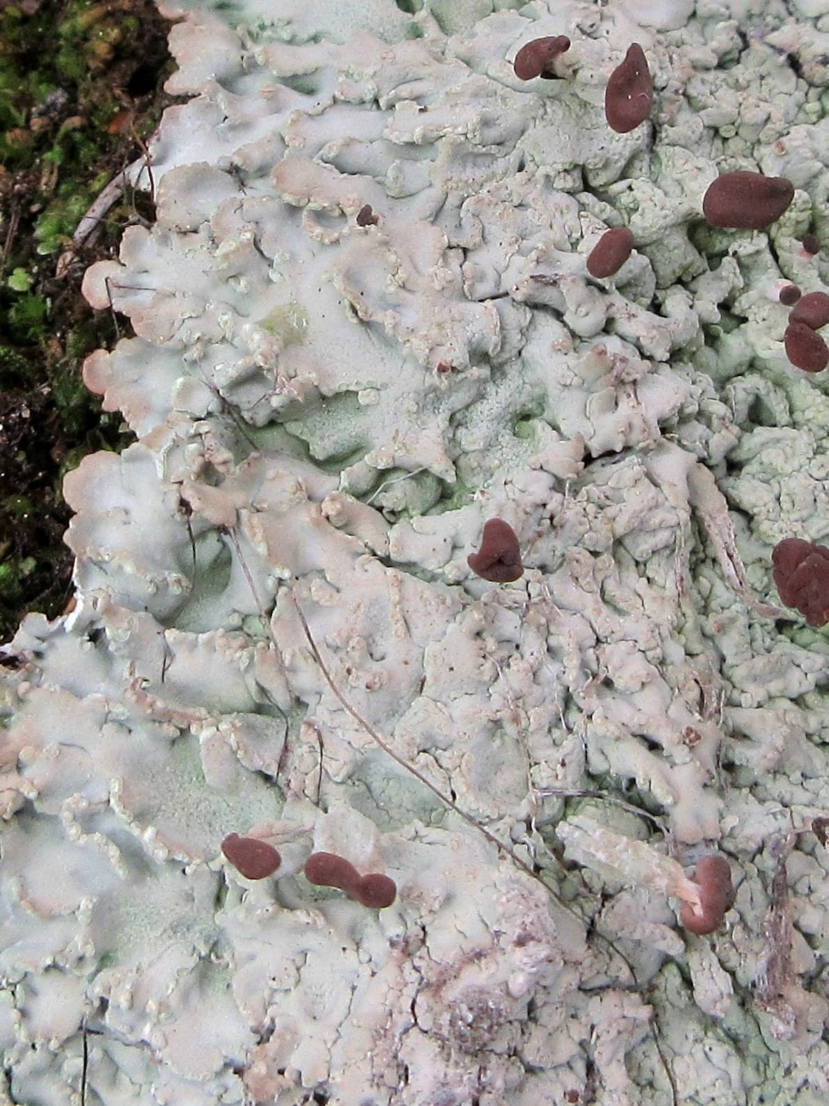
Baeomyces
Family: Baeomycetaceae
Baeomyces image
Troy McMullin
  • Greater Sonoran Desert
  • Resources
Nash, T.H., Ryan, B.D., Gries, C., Bungartz, F., (eds.) 2002. Lichen Flora of the Greater Sonoran Desert Region. Vol 1.
Life habit: lichenized Primary thallus: crustose, granular, verrucose or subsquamulose to squamulose, or marginally almost foliose upper surface: usually brown, gray, or olivaceous, ± continuous; soralia sometimes present; schizidia when present discoid, detachable cortex: with one or more pseudoparenchymatous layers or of interwoven hyphae running ± parallel to the upper surface photobiont: primary one a chlorococcoid alga, secondary photobiont absent; forming a zone at least in the primary thallus lower surface: without special structures Secondary thallus: composed of short stipes (mostly under 2 cm tall), erect, usually unbranched, solid; Ascomata: apothecial, borne terminally, 1-several, terminal on stipes, or sometimes almost sessile; disc: pale to dark brown or reddish brown (rarely pinkish), roundish, concave to flat and marginate at first, later swollen and with reflexed margins, often clustered, usually (at least the clusters) distinctly larger than diam. of the stipe; thalline exciple: absent asci: narrow, unitunicate, thin-walled, the apex truncated, with a single functional wall layer, I-, K/I-, 8-spored ascospores: ellipsoid, obtuse at the poles, simple to indistinctly 1-septate, without an endospore thickening, 8-14 x 2-4 µm Conidiomata: pycnidial, immersed in small warts on the thallus conidia: short, bacilliform Secondary metabolites: ß-orcinol depsidones Geography: cosmopolitan, circumpolar, arctic to tropical regions and Australasia Substrate: acidic rocks or soils, bryophytes, detritus; characteristic of temporary and recently disturbed sites. Notes: The genus is usually easily recognized (when fertile) by the ± pale brown, often convex and swollen apothecia usually on rather short and slender, solid stipes, arising from ± crustose (to minutely squamulose or lobate) thallus, growing on soil or rock. It differs from Cladonia especially in that the stalks are solid and its basal squamules, if present, are mostly tightly appressed and often fused. If the stipes are absent, the genus might be confused with various other crustose genera with ± pale biatorine apothecia. Species with distinctly pinkish apothecia, an amyloid hymenium, a different ascal structure, and containing depsides rather than depsidones, have now been placed in the genus Dibaeis. The statement by Thomson (1984) that B. roseus (= Dibaeis baeomyces) is the type species of the genus Baeomyces is now incorrect, because that species is the genus type for Dibaeis.
Show all taxa || << 1 - 50 taxa >>
Baeomyces acicularis
Images
not available
Map not
Available
Baeomyces aggregatus
Image of Baeomyces aggregatus
Map not
Available
Baeomyces auratus
Images
not available
Map not
Available
Baeomyces bacillaris
Images
not available
Map not
Available
Baeomyces botryophorus
Images
not available
Map not
Available
Baeomyces brevis
Image of Baeomyces brevis
Map not
Available
Baeomyces caesiopruinosus
Images
not available
Map not
Available
Baeomyces calicioides
Images
not available
Map not
Available
Baeomyces callianthus
Image of Baeomyces callianthus
Map not
Available
Baeomyces capitatus
Images
not available
Map not
Available
Baeomyces caprinus
Images
not available
Map not
Available
Baeomyces carneus
Image of Baeomyces carneus
Map not
Available
Baeomyces chilensis
Images
not available
Map not
Available
Baeomyces cinnabarinus
Image of Baeomyces cinnabarinus
Map not
Available
Baeomyces clauzadei
Images
not available
Map not
Available
Baeomyces crenulatus
Image of Baeomyces crenulatus
Map not
Available
Baeomyces cupreus
Images
not available
Map not
Available
Baeomyces demangei
Images
not available
Map not
Available
Baeomyces elveloides
Images
not available
Map not
Available
Baeomyces ericetorum
Image of Baeomyces ericetorum
Map not
Available
Baeomyces frenchianus
Image of Baeomyces frenchianus
Map not
Available
Baeomyces fuscocarneus
Images
not available
Map not
Available
Baeomyces fuscorufescens
Images
not available
Map not
Available
Baeomyces gomphillaceus
Images
not available
Map not
Available
Baeomyces granosus
Images
not available
Map not
Available
Baeomyces haemotropus
Images
not available
Map not
Available
Baeomyces helveloides
Images
not available
Map not
Available
Baeomyces heteromorphus
Image of Baeomyces heteromorphus
Map not
Available
Baeomyces hyalinus
Images
not available
Map not
Available
Baeomyces insignis
Images
not available
Map not
Available
Baeomyces jamesonii
Image of Baeomyces jamesonii
Map not
Available
Baeomyces lignorum
Images
not available
Map not
Available
Baeomyces marginalis
Image of Baeomyces marginalis
Map not
Available
Baeomyces novae-guineae
Images
not available
Map not
Available
Baeomyces novae-zealandiae
Images
not available
Map not
Available
Baeomyces novae-zelandiae
Images
not available
Map not
Available
Baeomyces pachypus
Image of Baeomyces pachypus
Map not
Available
Baeomyces paeminosus
Images
not available
Map not
Available
Baeomyces pertenuis
Images
not available
Map not
Available
Baeomyces pilatensis
Images
not available
Map not
Available
Baeomyces placophyllus
Image of Baeomyces placophyllus
Map not
Available
Baeomyces plagiocarpus
Images
not available
Map not
Available
Baeomyces prostii
Images
not available
Map not
Available
Baeomyces puiggarii
Images
not available
Map not
Available
Baeomyces pulogensis
Image of Baeomyces pulogensis
Map not
Available
Baeomyces rhodochrous
Images
not available
Map not
Available
Baeomyces rubescens
Images
not available
Map not
Available
Baeomyces rubescescens
Images
not available
Map not
Available
Baeomyces rufus
Image of Baeomyces rufus
Map not
Available
Baeomyces rupestris
Image of Baeomyces rupestris
Map not
Available


This project made possible by National Science Foundation Awards: #1115116, #2001500, #2001394
Powered by Symbiota