
Consortium of Lichen Herbaria
- building a Global Consortium of Bryophytes and Lichens as keystones of cryptobiotic communities -
- Home
- Search
- Images
- Species Checklists
- US States: O-Z >
- US National Parks
- Central America
- South America
- US National Parks
- Southern Subpolar Region
|
Baeomyces rufus (Hudson) Rebent.
 |
|
|
Family: Baeomycetaceae
Brown Beret Lichen
[Baeomyces byssoides (L.) Gärtner, Mey. & Schrebius, moreBaeomyces rufus f. subsquamulosus (Nyl.) Frey, Baeomyces rufus var. prostii Harm., Baeomyces rufus var. subsquamulosus Nyl., Lichen rufus Huds.]  Samuel Brinker |
Nash, T.H., Ryan, B.D., Gries, C., Bungartz, F., (eds.) 2002. Lichen Flora of the Greater Sonoran Desert Region. Vol 1. Primary thallus: discrete or ± wide-spreading, continuously crustose, nodulose, warty or partly squamulose, the squamules small (to 1 mm broad), somewhat raised, compacted, sometimes imbricate, ± delimited surface: green to dull greenish gray, to whitish gray or sometimes brownish, either not sorediate or with diffuse, coalescing, irregular greenish soralia with powdery soredia; schizidia occasionally present, < 0.2 mm diam., disc-like cortex: lacking where sorediate, composed of three types: 1) mainly in ± vertical paraplectenchyma, 2-3 cells thick or 2) composed of hyphae spreading out from between the algae and becoming ± flat-interwoven, or 3) forming a ± vertical but decomposed layer that is macroscopically whitish and pruinose photobiont: cells 6-13 (-14) µm diam., globose or a few ellipsoid, < 14 x 12 µm stipes: short, rarely more than 6 mm tall, flattened or cylindrical, white, furrowed and almost always fissured, mostly ecorticate but sometimes greenish and corticate towards the base; inner layer: composed of parallel thick-walled hyphae and containing many algal cells Apothecia: almost sessile, dark red-brown, seldom reddish or pale dull pink, ± translucent when wet, concave or flat, becoming slightly convex, often incurved at base, up to 2 mm diam., single or occasionally several together; exciple: distinct from the hypothecium, at edges gradually merging into a palisade plectenchyma; hymenium: 90-105 µm; paraphyses: slender, 1-1.8 µm, the tips scarcely thickened asci: cylindrical, 75-100 x 7-9 µm, I-, KI-, 8-spored ascospores: ellipsoid, often indistinctly 1-septate, 8-13 x 2.5-4.5 µm Pycnidia: often absent conidia: ellipsoid, 4-5 x 1 µm Spot tests: thallus K+ yellow, C-, KC+ yellow, P+ yellow to orange, UV- Secondary metabolites: stictic acid (major), norstictic and constictic acids (both accessory). Substrate and ecology: on soil in coniferous forests and particularly in recently disturbed, ± moist sites World distribution: circumpolar in temperate and boreal zones in the Northern Hemisphere, extending southwards in the mountains Sonoran distribution: eastern Arizona, 2900 m, on shaded earth in Picea engelmannii forest. Notes: Although it is a distinctive species, mature specimens have not yet been found in the Sonoran region. It was recognized by Dr. J. Hafellner on the basis of its parasite occurring on its crustose thallus. |
 Troy McMullin 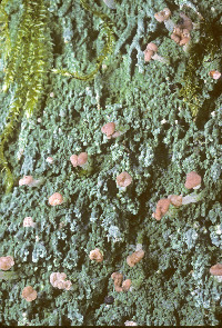 Stephen Sharnoff  Stephen Sharnoff 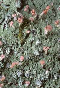 Stephen Sharnoff 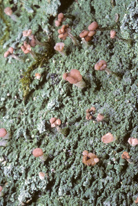 Stephen Sharnoff  Stephen Sharnoff  Stephen Sharnoff 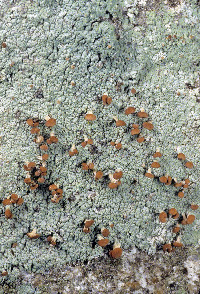 Stephen Sharnoff  Stephen Sharnoff 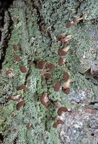 Stephen Sharnoff  Lucy Taylor  ") ") ") ") ")               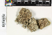   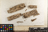   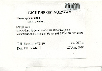 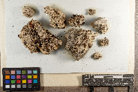  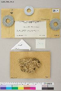  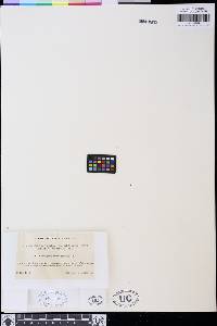     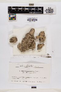          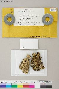  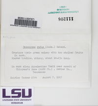  ")       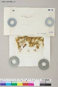                   |
|

![]()
![]()

![]()
![]()
![]()
![]()
![]()
Powered by Symbiota