Acarospora succedens
|
Acarospora succedens H. Magn.
 |
|
|
Family: Acarosporaceae
[Acarospora interspersa H. Magn.]  |
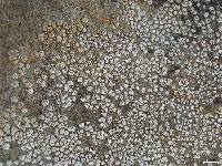
Acarospora succedens H. Magn., Meddn Göteb. Bot. Trädg. 5: 71 (1930) [1929]. Type: USA. New Mexico: Environs de Las Vegas, Arsène Brouard 19950 (Hb. B de Lesdain, holotype, lost in WW2, n.v. FH! isotype). MB#375738 = Acarospora interspersa H. Magn., Annals Cryptog. Exot. 6(1): 45 (1933). Type: USA. New Mexico: Environs de Las Vegas, Canon Sud, Arsène Brouard 20557 (UPS! holotype). MB#410163 Taxonomic Note: This is a newly updated description (see also taxonomic comments added to previous descriptions in the Lichen Flora of the Greater Sonoran Desert Region). Description. Hypothallus endosubstratal, no algae observed. Thallus areolate, to 5 cm wide; areoles round to irregular, flat to convex, (0.2–)1.0(–1.3) mm in diam., 0.2–0.6 mm thick, solitary, dispersed, or emerging along an axis from cracks in the rock, or morphing out of other lichens, becoming contiguous through replication by division. Upper surface dark to yellow brown or an orange-brown, sometimes glossy, epruinose, rugulose, densely fissured in mature areoles, smooth in young areoles, the fissures beginning often as small pits. Epicortex 6–35 µm thick, with thin periclinal to intricate hyphae usually visible. Upper and lateral cortices paraplentenchymatous but originating from anticlinal hyphae, 30–40 µm thick, upper layer various shades of golden or reddish brown fading into indistinct lower hyaline zone 15–20 µm thick. Algal layer uneven, algal cells not dense, interrupted by hyphal bands (but in very flat specimens appearing solid with plane upper surface). Medulla white, obscure, intricately prosoplectenchymatous. Lower surface white, ecorticate, narrow around the mycelial base of the areole which is broadly attached to substrate, continuous with the hypothallus and the medulla, and eventually elevating the areole without forming a stipe (gomphate). Apothecia 1–4 per disc, forming in the center of areole, immersed, with visible beginning of a thalline margin; rarely the immersed apothecium reduce the areole to a thalline margin. Disc brown or black, plane, smooth, or often rugulose with plectenchyma forming gyrose formations. Parathecium expanding around disc to c. 35 µm wide. Hymenium pale yellow to hyaline, 80–110 µm tall, epihymenium reddish brown, thickly conglutinated, 15–20 µm thick, paraphyses 1.5–2.0 µm wide at base, apices expanded to 3–4 µm wide, hymenial gel IKI+ blue turning red, hemiamyloid. Asci narrowly clavate, 70 × 10–15 µm, 100+ ascospores hyaline, simple, ellipsoid (3–)4.5–6 × 1.5–2 µm. Subhymenium c. 10 µm thick, IKI+ dark blue. Hypothecium distinct, c. 10 µm thick. Pycnidia c. 100 µm in diam., conidia 2.5–3.0 × 0.8–1 µm (Magnusson 1933). Chemistry. Secondary metabolites with HPLC (Knudsen 2007): gyrophoric acid (major), lecanoric acid (minor), 3-hydroxygyrophoric acid (trace), methyl lecanorate (trace). Spot tests: cortex KC+ C+ red. Note. Acarospora succedens differs from all these species with gyrophoric-lecanoric acid in Southwestern North America in having densely fissured yellow-brown to dark brown areoles wth thick mycelial base. None of the other species in the Southwest producing gyrophoric/lecanoric acid are also facultative lichenicolous lichens. The small pits, which are the beginning of fissuring, on usually small sterile areoles can be confused with A. obpallens, but the fertile and fissured mature areoles are unmistakable as A. succedens. Magnusson reports an unusually long conidia length for an Acarospora (Magnusson 1933). We have not been able to verify it. Nash, T.H., Ryan, B.D., Gries, C., Bungartz, F., (eds.) 2007. Lichen Flora of the Greater Sonoran Desert Region. Vol 3. Important Taxonomic Note: The description below was originally published for Acarospora interspersa in the Lichen Flora of the Greater Sonoran Desert Region. When isotype material of A. succedens was discovered at FH, it became evident that the previous concept of that taxon had been erroneous and that the name instead had to be applied to A. interspersa, which accordingly is now considered a synonym of A. succedens (Knudsen 2011). The description previously published for A. succedens instead applies to a species newly described by Kudsen (2011) as A. nashii (see notes there). Thallus: areolate, overall up to 5 cm wide areoles: usually round, (0.2-)0.7-1(-1.3) mm in diam., 0.2-0.6 mm thick, solitary, dispersed or emerging along an axis or from other lichens, becoming contiguous through division of the thallus along fissures in upper surface down through attaching hypha; rim: ±down-turned, sometimes white upper surface: dark to yellow brown or an orange-brown, glossy, convex, rugulose, fissures evident in even smallest thallus, epruinose lateral cortices: paraplectenchymatous but originating from anticlinal prosoplectenchyma, 30-40 µm thick; cells: regular, obscured in water, clear in K; syncortex: 6-35 µm thick, with thin periclinal to intricate hyphae usually visible; eucortex: upper layer various shades of golden or reddish brown fading into indistinct lower hyaline zone 15-20 µm thick medulla: white, obscure, intricately prosoplectenchymatous lower surface: white, ecorticate algal layer: uneven, algal cells not dense, interrupted by hyphal bands (but in very flat specimens appearing solid with a plane upper surface) attachment: broad and eventually elevating areoles (gomphate) without forming a stipe Apothecia: 1-4 per disc, forming in the center of areole, immersed, with visible beginning of a thalline margin, rarely becoming sessile and fully lecanorine disc: brown or black, plane, smooth or rough, sometimes convex; occasionally with interascal plectenchyma forming gyrose formations parathecium: expanding around disc up to c. 35 µm wide epihymenium: reddish brown, thickly conglutinated, 15-20 µm thick hymenium: pale yellow to hyaline, 80-110 µm tall; paraphyses: 1.7-2 µm wide at base; apices expanded, 3-4 µm wide subhymenium: pale yellow, c. 10 µm thick; hypothecium: distinct, c. 10 µm thick asci: narrowly clavate, 70 x 10-15 µm, 100+-spored ascospores: hyaline, simple, narrowly ellipsoid (3-)4.5-6 x 1.6-2 µm Pycnidia: c. 100 µm in diam. (Magnusson 1933) conidia: bacilliform, 2.5-3 x 0.8-1 µm (Magnusson 1933) Spot tests: UV-, cortex C+ red Secondary metabolites: gyrophoric acid (major), lecanoric acid (minor), 3-hydroxygyrophoric acid (trace), methyl lecanorate (trace) (HPLC, J.A. Elix, pers comm.) Substrate and ecology: on granite and volcanic rock; juvenile parasite in one specimen (Nash 16, 215 ASU) growing on and out of Aspicilia species, but no evidence it is obligate or host-specific parasite World distribution: southwestern North America and Mexico Sonoran distribution: Arizona, southern California (Santa Cruz Island), Baja California (Guadalupe Island), Baja California Sur and Sonora. Notes: This description differs slightly from details of Magnusson's description of the holotype (UPS!), because the type is not a typical specimen. The center of distribution appears to be in Mexico. Specimens differ and a more accurate description and distribution should emerge from further collections and study. Some specimens of A. nevadensis can look similar to A. interspersa, but the areoles of the former species are not as consistently round and its spores are broadly ellipsoid and have a mucilaginous layer around the thick walls. Tentatively a collection by Wet-more with no chemistry from Santa Cruz Island is included in this taxon; its spot tests reactions are negative, and it needs further comparisons with collections from the Channel Islands and central California. Relevant Literature: |


")