
Consortium of Lichen Herbaria
- building a Global Consortium of Bryophytes and Lichens as keystones of cryptobiotic communities -
- Home
- Search
- Images
- Species Checklists
- US States: O-Z >
- US National Parks
- Central America
- South America
- US National Parks
- Southern Subpolar Region
|
Arthonia sanguinea Willey
 |
|
|
Family: Arthoniaceae
[Arthothelium sanguineum (Willey) Zahlbr.] 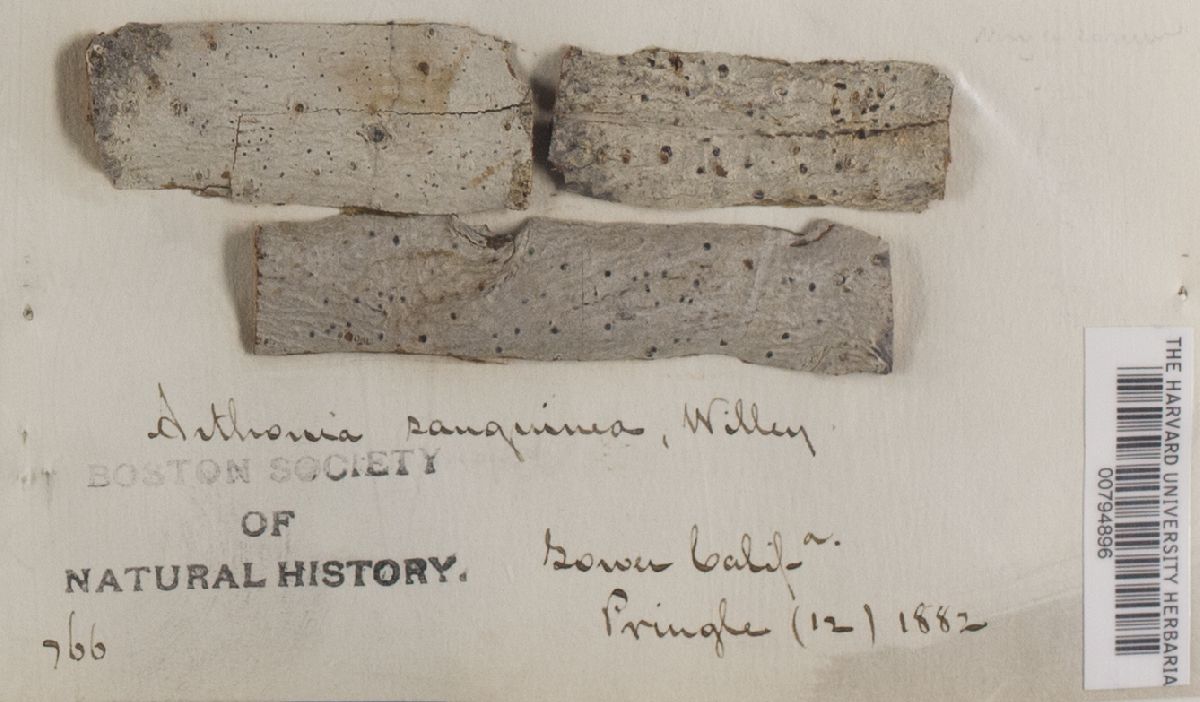 |
Nash, T.H., Ryan, B.D., Gries, C., Bungartz, F., (eds.) 2007. Lichen Flora of the Greater Sonoran Desert Region. Vol 3. Thallus: ±white, effuse, inconspicuous photobiont: not seen Ascomata: ±round to irregularly elongate, 0.48-0.72 mm in diam., in section 100-140 µm tall, ±at the same level as thallus disc: black, occasionally becoming strongly convex (and then bark disrupted below the subhymenium), with dark red pruina; exciple: similar to epithecial structures, often more green, rarely with yellowish green to orange pigment particles epihymenium: pale to dark brown, 20-30 µm thick, with fine granular or patches of reddish pigments; hyphal structure: similar as in the layers below, but hyphae more branched; cells: 4-8 x 2-3 µm, with olivaceous brown walls hymenium: ±hyaline to pale yellow, 60-70 µm tall; paraphysoids: branched and anastomosing, embedded in a rigid gel, not concentrated around the asci; cells: 4-8 x 1-2 µm; asci widely spaced; subhymenium: hyaline to yellowish brown, 20-40 µm thick; cells: 3-5 µm large asci: subglobose, 47-60 x 35-55 µm, 8-spored, distinctly stipitate, lateral endotunica thickened ascospores: hyaline, muriform, with 5-7 transverse septa and 1-3 longitudinal septa in each transverse segment, ±ellipsoid, (19)23-30 x (9)10-20 µm Pycnidia: not observed Chemical reactions: ascomatal gels Idil+ deep blue, I+ deep blue (even thin slices opaque), KI+ blue; epihymenial red pigments K+ pale brown but not dissolving.; asci without KI+ reactive tholus structures. Substrate and ecology: on lignified substrates (e.g., Tamarix, Euphorbia), not lichen-forming World distribution: Mexico (Baja California), Africa (Senegal), and Asia Minor (Cyprus) Sonoran distribution: southern California (Santa Barbara and Catalina Island), Baja California, and Baja California Sur. Notes: Arthonia sanguinea has repeatedly been confused with Arthothelium spectabile. Superficially, the ascomata of Arthonia sanguinea resemble those of Arthothelium s. str. However, the structure of the paraphysoids is clearly different and the species cannot be accommodated in the same species group. The paraphysoids are not conspicuous in a transmission light microscope, but they can be well observed after application of K, or in epifluorescence microscopy after staining with calcofluor white. Vertically arranged elements predominate in A. sanguinea, and they do not entangle the asci densely, as in A. spectabile, which also differs in having a more compact gelatinous matrix. Arthonia sanguinea is known on different continents but always from sites influenced by coastal fogs. The specimen from Africa has slightly smaller ascospores and ascomata, which are not as distinctly convex as in the American specimens. Other characters, such as the pigmentation of the ascomata are identical in other locations. The species was rarely collected in the past but seems to be one of the very common species in the Sonoran area. |
 Chris Wagner  Chris Wagner  Chris Wagner  Chris Wagner ") ")  Ken Kellman 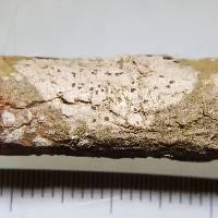 Gary Perlmutter ") ")  ") ") 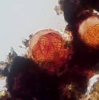 Gary Perlmutter ") ") 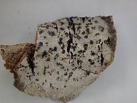 Ken Kellman 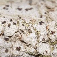 Gary Perlmutter ") ")  Chris Wagner  Gary Perlmutter ") ") ") 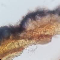 Gary Perlmutter ") ")  Gary Perlmutter  Ken Kellman |
|

![]()
![]()

![]()
![]()
![]()
![]()
![]()
Powered by Symbiota